Scientia et Arte
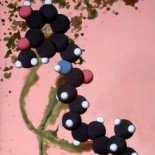
If science were art and art were science, then the howling black wolf has probably swallowed some N-(4-hydroxyphenyl)ethanamide.
Wilson Disease
Wilson disease is an autosomal recessive inherited disorder of copper metabolism. It is primarily characterized by excessive copper deposits in liver, kidney, and brain. It was first described by Dr. Samuel Alexander Kinnier Wilson, a British neurologist, in 1912 and was named after him.

Copper Metabolism
An important trace mineral in the body (1-2mg daily intake), copper serves as cofactor for several mammalian enzymes, such as cytochrome c oxidase (mitochondrial respiration), lysyl oxidase (collagen and elastin cross-linking), tyrosinase (melanin production) and ceruloplasmin (copper exportation from liver). It also functions as catalyst because of its distinct redox states. It plays a role in immune response, as well. Furthermore, copper metabolism is closely linked with iron metabolism.
In normal copper metabolism, most of the copper that enters the body is excreted in urine and stools. For patients with Wilson’s disease, copper metabolism in enterocytes, small intestine cells, are normal; however, in hepatocytes, liver cells, there is a markedly decreased copper excretion in bile, ceruloplasmin, and stools. (See Figure 1.2 and 1.3 for comparison of normal and abnormal copper metabolism.)
.gif)

As copper enters the digestive tract, about 25% of its total amount is directly excreted in the stools. Fifty-percent is absorbed into the enterocytes and complexes with metallothionein, protein that binds metals and stores them in non-toxic form. Then, ATP7A (copper-transporting ATPase1), a copper transporter protein in enterocytes, delivers albumin-complexed copper into the liver. (Detailed cellular hepatic metabolism is illustrated in Figure 1.4.) In the enterocytes, copper is directed by ATOX1 chaperone protein to metallothionein and ceruloplasmin. Metallothionein stores copper intracellularly and the excess is excreted into ATP7B-regulated bile capillaries. Ceruloplasmin, a six-copper binding 2-globulin, is then formed from the ATP7B-regulated transfer of copper to apoceruloplasmin. Finally, it circulates in the blood and delivers copper to brain and kidneys, as the major transport protein copper carrier.

Wilson disease involves the mutation chromosome 13q14.3–q21.1 wherein the ATPase, Cu2+-transporting, beta polypeptide (ATP7B) gene is located (Fig. 1.5). Consisting of 1 411 amino acids, the gene encodes P-type ATPase protein, the primary protein in copper homeostasis in the liver. ATP7B protein consists of an ATPase domain, a hinge domain, a phosphorylaiton site, and six-copper binding sites (Fig. 1.6). In afflicted patients, low levels of expression of the gene predominantly in the liver, but also in the kidneys, brain, lungs, and heart are identified. This deficiency results to increased apoceruloplasmin levels, decreased ceruloplasmin levels, and toxic build-up of copper in hepatocytes. As excess copper collects in the liver, mitochondria are damaged and copper leaks into the blood which eventually collects in the brain, kidneys, eyes and lungs. They promote free radical formation that results in oxidation of lipids and proteins.


Symptoms

Because Wilson Disease is characterized by the accumulation of copper in the body, its symptoms are not evident during birth but only manifest after 20-30 years. They include mostly neurological, hepatic, renal, psychiatric, and haematological signs. As copper collects in the brain, cognitive deterioration, psychosis, depression, behavioral changes, seizures and migraine occur. Hepatic symptoms include cirrhosis, acute hepatitis, jaundice, and liver failure. Upon slit lamp examination, golden-brown deposits are visible in the cornea. These are called Kayser-Fleischer rings (Fig 1.8). It is also characterized by the excretion of the normally absorbed substances by the urine. Hemolysis is also observed as red blood cells are damaged by the leaked copper in bloodstream.
Treatment
Patients with Wilson Disease are initially treated through chelation with D-penicillamine and trientine. Their by-products bind with copper and excrete it through the urine. Zinc in the form of zinc acetate, is also prescribed. It promotes binding of copper to metallothionein in the intestines. Copper is eventually excreted in stools and is prevented to travelling to the liver. In some cases, when the liver is unresponsive to chelation, liver transplants are conducted.
References:
http://jn.nutrition.org/content/133/5/1527S.full
http://mounier.univ-tln.fr/rcmo/php_biblio/PDF/5293.pdf
http://www.eurowilson.org/en/living/guide/pathway/index.phtml
http://www.nature.com/nrneurol/journal/v2/n9/full/ncpneuro0291.html
[post by Kathee Prado]
